When Whitening Meets Restorations: How Bleaching Affects Tooth-Colored Fillings
Bright Smiles, Fading Fillings?
Tooth bleaching has become one of the most popular cosmetic dental procedures worldwide. Whether performed at home or in the clinic, whitening treatments promise brighter smiles — but what happens to existing dental restorations when exposed to these bleaching agents?
A research team from the University of Malaya set out to explore exactly that: how whitening products influence the color stability of modern ion-releasing restorative materials — the tooth-colored fillings designed to release beneficial minerals and resist recurrent caries.
Background: Smarter Fillings for Healthier Teeth
Traditional composite resins are prized for their beauty and strength, but they’re not perfect. Over time, the edges of restorations can attract plaque and lead to secondary cavities.
To combat this, ion-releasing restoratives (IRMs) were developed. These materials release fluoride, calcium, and phosphate ions that can help remineralize teeth and prevent decay. However, their performance under bleaching conditions — especially newer types like bioactive composites and giomers — has remained uncertain.
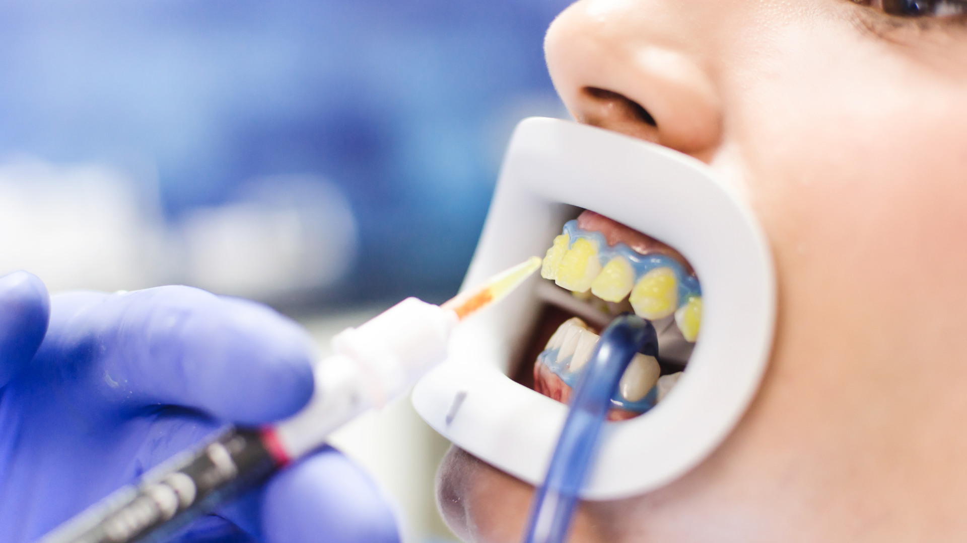
The Study: Putting Restoratives to the Test
Researchers evaluated five types of restorative materials:
Activa Bioactive (bioactive composite)
Beautifil II LS (giomer)
Cention-N (alkasite)
Riva LC HVGIC (resin-modified glass ionomer)
Luna (conventional composite resin, used as control)
Each material was shaped into small discs and exposed to one of three conditions:
In-office bleaching (using Pola Rapid, 38% hydrogen peroxide)
At-home bleaching (using Pola Night, 22% carbamide peroxide)
Control group (stored in artificial saliva only)
Color changes were measured over one month using a spectrophotometer based on the international CIE-Lab color system — a method that quantifies changes in lightness and hue.
Key Findings: Material Matters
Bleaching does change the color of restorations — but not equally across materials.
Riva LC HVGIC showed the greatest color change immediately after bleaching, while Luna remained the most stable.
Activa Bioactive continued to discolor over time, showing the largest overall color shift after one month.
Interestingly, both in-office and take-home whitening systems produced similar effects on all materials.
The study also found that even artificial saliva alone could cause color changes in some restoratives — showing that discoloration is not just about bleaching but also long-term material behavior.
What This Means for Dentists and Patients
Color changes may seem subtle to the untrained eye, but once the difference (ΔE*) exceeds a threshold of around 2.7, it becomes clinically visible and potentially unacceptable.
For patients, this means:
Whitening treatments can alter the appearance of existing fillings.
Some bioactive and glass ionomer-based materials may need replacement after bleaching to match tooth color.
For clinicians:
Restorative procedures should ideally be postponed at least one week after bleaching to allow color stabilization.
Choosing materials with proven color stability — such as giomers or alkasites — can minimize esthetic issues.
Study Limitations and Future Directions
The experiment used controlled lab conditions, not real mouths. Factors like saliva proteins, biofilm buildup, and temperature changes were not replicated. The authors recommend future studies incorporating these elements and using more advanced color measurement methods (like the CIEDE2000 system) for greater clinical accuracy.
Conclusion
This study highlights that while tooth whitening is safe and effective, its impact on dental restorations depends on the material type. Patients should discuss potential color mismatches with their dentists before undergoing bleaching, and clinicians should carefully select restorative materials when planning esthetic treatments.
Reference:
Lee, J.S., Yahya, N.A., Abdul Aziz, A., & Yap, A.U.-J. (2025). The effects of bleaching products on the color stability of ion-releasing restoratives. BMC Oral Health, 25, 1754.
DOI: 10.1186/s12903-025-06681-0