When Angles Matter: How Implant Tilt Affects Digital Scanning Accuracy in Dentistry
Digital Dentistry Meets Precision
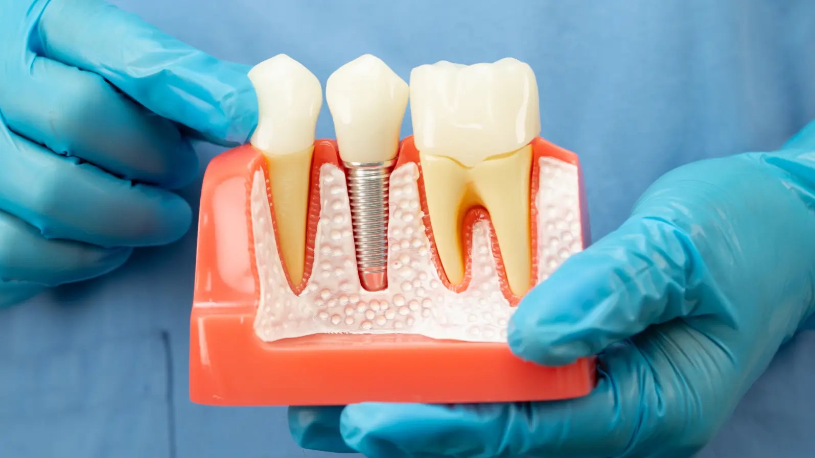
Digital tools are transforming modern dentistry — from diagnosis to the creation of perfectly fitted implant restorations. Among these, intraoral scanners (IOS) and photogrammetry (PG) have become central to creating accurate digital impressions of the mouth.
However, when implants are placed at an angle — often necessary due to bone structure or anatomy — the accuracy of these digital scans can be compromised. To explore how implant angulation affects scanning accuracy, researchers Tezcan Muslu and Sevcan Kurtulmus-Yilmaz from Near East University, Türkiye, conducted a detailed laboratory study comparing different scanning methods and auxiliary device designs.
The Study: Testing Angles and Devices
The researchers built three upper jaw models with five implants each, mimicking real clinical cases:
Model 1: All implants were parallel.
Model 2: One implant tilted 25° mesially (toward the midline).
Model 3: One implant tilted 25° distally (away from the midline).
Five digital impression techniques were compared:
IOS (basic intraoral scanning)
IOS-C (IOS with crown-shaped prefabricated auxiliary device, or PAD)
IOS-LE (IOS with laterally extended PADs)
PG-I (photogrammetry using ICam4D G3 system)
PG-O (photogrammetry using OxoCore system)
PADs are small 3D-printed attachments placed over scan bodies to help the scanner identify reference points more reliably — like adding “landmarks” for smoother digital stitching.
Key Findings: Not All Scans Are Equal
The results revealed clear performance differences:
The PG-I system (ICam4D G3) showed the highest accuracy, with minimal error (around 18 µm), remaining stable across all implant angles.
The IOS-LE technique — intraoral scanning with laterally extended PADs — also performed impressively, showing accuracy comparable to the photogrammetry systems.
Standard IOS scans without PADs were the least accurate, especially when implants were tilted distally.
Interestingly, implant angulation affected “trueness” (how close the scan is to reality) but not precision (how consistent the results are).
In simple terms:
➡️ Angled implants make it harder for scanners to “see” accurately, especially when tilted backward.
➡️ Adding laterally extended PADs or using photogrammetry helps maintain accuracy, even under these challenging conditions.
Why It Matters for Dentists
In full-arch implant restorations — such as complete upper or lower dentures — achieving a “passive fit” (no stress or misalignment when the prosthesis is placed) is essential for long-term success.
This study highlights that photogrammetry and PAD-assisted intraoral scanning can help dentists capture accurate digital impressions, even when implants are angled.
It also suggests that 3D-printed auxiliary devices (like the low-cost PLA designs tested here) could become practical tools for improving digital scanning outcomes without adding major complexity or cost.
Takeaway Message
For complex implant cases involving angulated implants:
Photogrammetry (PG-I) remains the most precise technique.
Intraoral scanning with laterally extended PADs (IOS-LE) is a promising, accessible alternative.
Both methods produce accuracy well within clinically acceptable limits.
In the fast-moving world of digital dentistry, this research reinforces that designing smarter scanning accessories and refining workflows can make advanced technologies more reliable and accessible for everyday practice.
Reference:
Muslu, T., & Kurtulmus-Yilmaz, S. (2025). Effect of implant angulation on the trueness and precision of intraoral scanning and photogrammetry. BMC Oral Health, 25, 1739. https://doi.org/10.1186/s12903-025-07107-7