When 3D-Printed Dental Tools Go Missing: Why They’re Hard to See on X-rays
A Hidden Problem in Digital Dentistry
Digital technologies have transformed modern dentistry. Today, surgeons can plan complex implant or jaw surgeries on a computer and create patient-specific guides with a 3D printer. These lightweight plastic templates help place implants precisely and reduce surgery time.
But there’s a catch: if a guide or template fractures during surgery—a rare yet possible event—tiny fragments could be swallowed, inhaled, or left inside the surgical field. Detecting those fragments quickly is crucial for patient safety. The question is: can current imaging systems actually see them?
The Study: Testing Visibility Under the X-ray Lens
Researchers from RWTH Aachen University and collaborating centers in Germany set out to test whether today’s commonly used 3D-printed dental materials are visible in standard radiologic scans.
They examined 15 materials frequently used for digital guides, models, and temporary restorations—ranging from well-known polymethyl methacrylate (PMMA) plastics to newer printable resins like MED610, PEEK, and Formlabs resins.
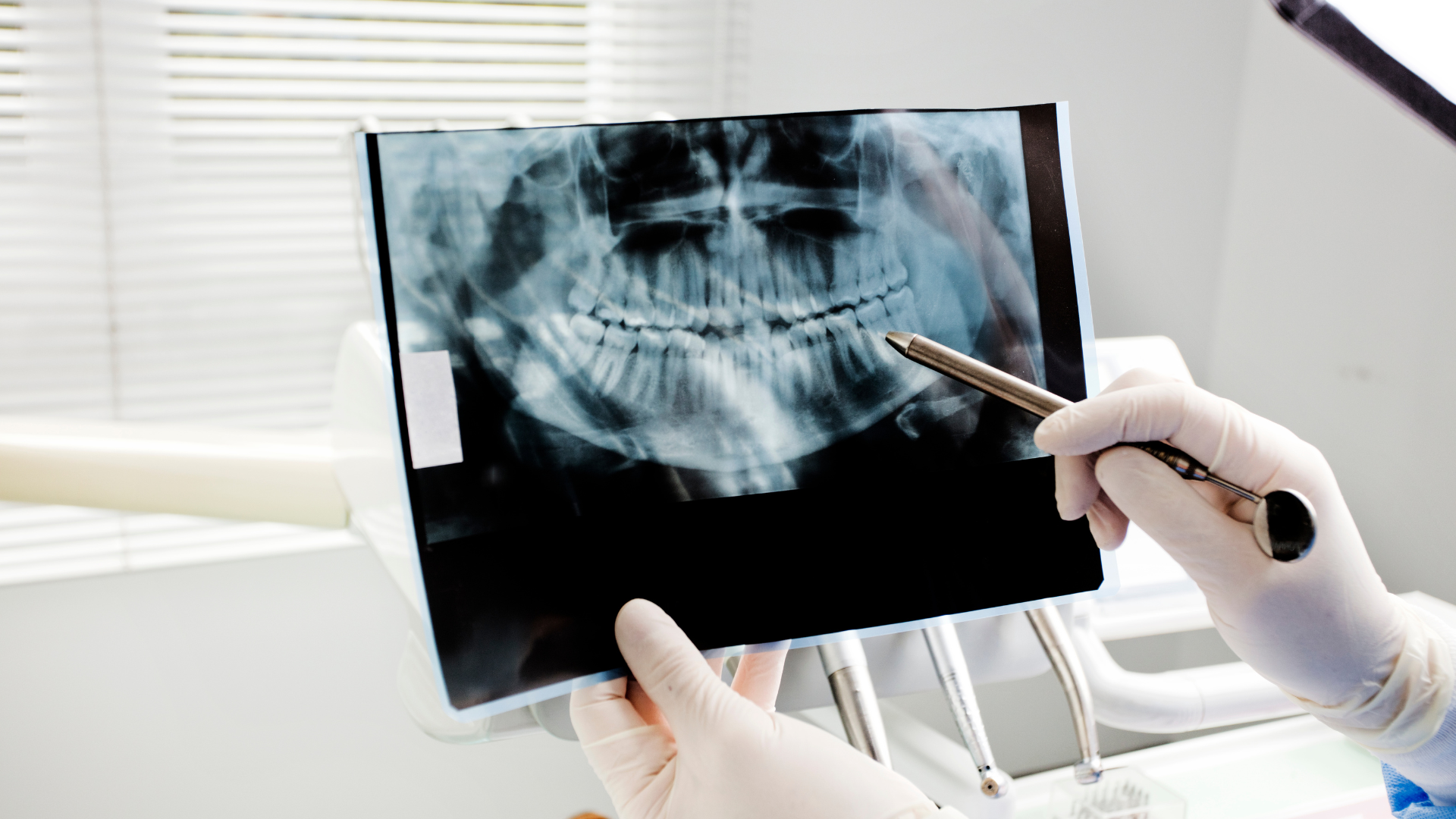
Using cone-beam computed tomography (CBCT) and conventional CT, the team compared how clearly these materials appeared when embedded in animal muscle and fat tissue—essentially simulating what happens if a fragment ends up inside a patient.
What They Found
Most 3D-printed materials were nearly invisible on both CT and CBCT scans.
Their radiodensity—measured in Hounsfield Units (HU)—ranged from about 69 to 130 HU, almost identical to that of soft tissues like muscle.
That means small fragments blended in so well with the surrounding tissue that even trained radiologists struggled to identify them.
Only traditional materials such as Luxatemp® and Futar D®, which contain naturally radiopaque components, stood out clearly on scans (with HU values above 1000).
In short: modern printable plastics look almost the same as flesh on X-rays.
Why It Matters
If a 3D-printed surgical guide or dental appliance breaks, a missing piece might escape detection, posing health and legal risks.
Unlike metals or ceramics, these newer polymers contain no heavy elements that absorb X-rays strongly. Without visible contrast, even high-resolution CBCT scanners—the gold standard in dentistry—may fail to reveal them.
The study also highlights a regulatory gap: while guidelines exist for mechanical safety and biocompatibility, no standards currently define minimum radiopacity for printable dental materials.
Possible Solutions
Researchers suggest improving safety by adding radiopaque fillers—for example, barium sulphate or triphenyl bismuth—to printable resins. These additives could make the materials more visible on scans without compromising print accuracy or strength.
Manufacturers could also publish each material’s imaging properties in product data sheets and contribute to an open database, helping clinicians choose safer options.
Until then, dentists are advised to design 3D-printed surgical tools with sufficient thickness and strength to minimize breakage and to handle them carefully during use.
The Takeaway
As 3D printing becomes routine in oral and maxillofacial surgery, ensuring the radiographic detectability of printed materials is an emerging safety priority.
This research serves as a timely reminder: in the quest for precision and innovation, what we can’t see might still matter most.
Reference
Niederau, C., Craveiro, R.B., Wolf, M., Becker, P., Pabst, A., & Zeller, A.-N. (2025).
Limited radiographic detectability of novel 3D-printed materials used in dental surgery.
BMC Oral Health, 25:1758. DOI: 10.1186/s12903-025-07158-w