Sharper Vision for Early Tooth Decay: Introducing the ACTA-DIRECT Micro-CT Dataset
A New Way to See What Dentists Often Miss
Early tooth decay especially proximal caries, which develop between teeth can be hard to spot. Even with dental X-rays, many early lesions simply don’t show up clearly enough. This can delay treatment, allowing small problems to grow into cavities that require drilling or restoration.
A new study from the Academic Center for Dentistry Amsterdam (ACTA) introduces a breakthrough resource that could change this:
the ACTA-DIRECT dataset, the first collection that pairs dental radiographs with highly detailed micro-CT scans to improve how researchers and AI systems detect early caries.
Why Early Caries Detection Is Challenging
Traditional radiographs are essential in dentistry, but they’re far from perfect. Studies show that early proximal caries have low detection sensitivity on radiographs down to 24–43% in some analyses. Much of the earliest enamel damage goes unnoticed.
Micro-CT imaging, on the other hand, offers extremely high-resolution 3D views of teeth. Though too impractical (and too high-radiation) for clinical use, micro-CT is ideal for creating research-quality reference standards. This helps eliminate variations in interpretation that typically occur between dentists.
What the Researchers Did
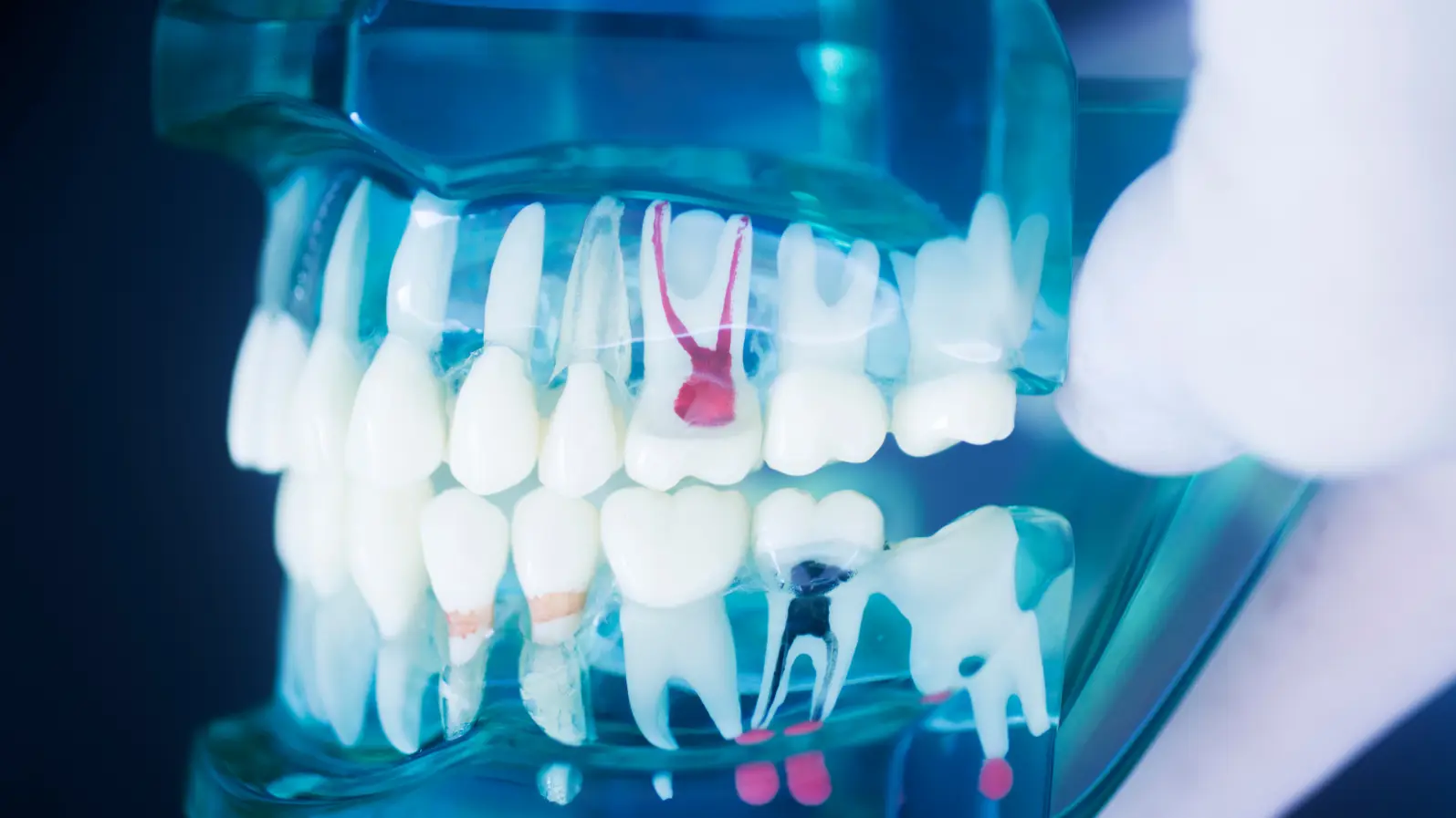
The ACTA team created a dataset of 179 extracted teeth, each scanned with:
Micro-CT (high-resolution 3D imaging)
Standard dental radiographs
Three types of caries annotations:
Conventional radiograph annotations
Micro-CT–assisted radiograph annotations
Micro-CT ground-truth annotations by an expert radiologist
Three dentists independently marked early caries on radiographs with and without assistance from the corresponding micro-CT scans. The team then compared accuracy, consistency, and agreement across methods.
Key Findings: Micro-CT Makes a Big Difference
The results were clear: micro-CT assistance substantially improved accuracy.
1. Better Agreement Between Dentists
Conventional radiographs: Cohen’s Kappa = 0.46
With micro-CT assistance: Kappa = 0.64
This shift from moderate to substantial agreement shows that micro-CT guidance reduces guesswork.
2. Higher Diagnostic Accuracy
Using micro-CT support:
Sensitivity rose from 42% → 63%
Specificity rose from 92% → 95%
Positive predictive value increased from 75% → 88%
ROC-AUC improved from 0.67 → 0.78
Dentists became significantly better at identifying early lesions and distinguishing sound surfaces from true caries.
3. Better Reference Data for AI Training
High-quality datasets are essential for developing trustworthy dental AI. Because traditional radiograph annotations have low sensitivity, AI models trained on them may inherit the same weaknesses.
The ACTA-DIRECT dataset offers the first micro-CT-based ground truth for early proximal caries, making it a valuable resource for advancing dental AI.
Why This Matters
Early caries are the ideal stage for preventive treatment no drilling, no fillings. But since these lesions are hard to detect, they often progress unnoticed.
By providing a precise, standardized dataset, ACTA-DIRECT can help:
Improve AI-based caries detection tools
Support research on early-stage lesion analysis
Enhance training of future dental diagnostic technologies
The dataset is publicly available and designed to grow in the future.
Conclusion
The ACTA-DIRECT dataset represents a major step forward in the quest for more accurate dental diagnostics. By combining micro-CT precision with conventional radiographs, the study shows how imaging innovations can improve early caries detection and ultimately support better patient care.
Original Article
Valenzuela RE, Mettes P, Loos BG, Marquering H, Berkhout E. Enhancement of early proximal caries annotations in radiographs: introducing the Diagnostic Insights for Radiographic Early-caries with micro-CT (ACTA-DIRECT) dataset. BMC Oral Health. 2024;24:1325.
DOI: https://doi.org/10.1186/s12903-024-05076-x