Seeing Inside the Root Canal Without a Scope
How CBCT-Based Virtual Endoscopy Is Opening a New Era in Digital Endodontics
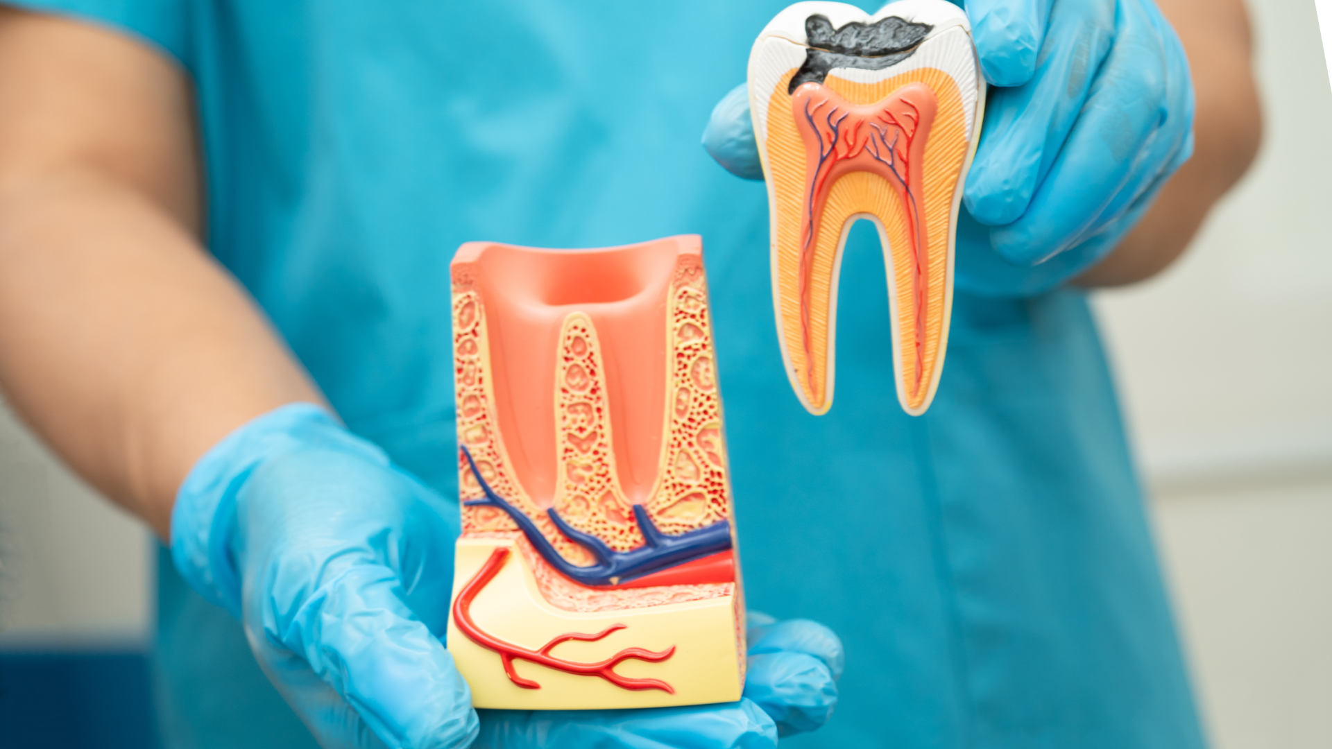
Opening: A New Way to “Look Inside” Teeth
Successful root canal treatment depends on one crucial factor: understanding the hidden anatomy inside the tooth. Yet, for decades, dentists have relied on two-dimensional X-rays to guide a three-dimensional procedure. Important anatomical details such as accessory canals or the exact position of the apical foramen can easily be missed.
A study published in the Brazilian Dental Journal introduces a breakthrough approach: a virtual root canal endoscopy created using advanced cone-beam computed tomography (CBCT) software. This innovation promises to change how clinicians visualize, plan, and communicate endodontic treatments.
Why Conventional Imaging Is Not Enough
Periapical radiographs remain the most common imaging tool in endodontics. However, they flatten complex structures into a single plane. Even standard CBCT scans, while more informative, may still limit the clinician’s ability to truly “see” inside the canal system.
The authors highlight that internal tooth anatomy often hides microstructures lateral canals, apical deltas, subtle curvatures that directly influence treatment outcomes. Missing these details can lead to persistent infection or treatment failure.
What Did the Researchers Do Differently?
The research team developed a computational modeling method that simulates endoscopy inside the root canal without inserting any physical endoscopic device.
Using ultra-thin CBCT slices (0.10 mm) and a specific 3D filter called the “pulp cavity filter” within the e-Vol DX CBCT software, they created a virtual environment where dentin becomes transparent while the pulp cavity remains visible.
This allows clinicians to:
Navigate dynamically inside the canal system
Switch seamlessly between 2D and 3D views
Visualize microanatomy as if traveling through the canal itself
In simple terms, the tooth becomes a digital replica that can be explored from the inside.
Real Clinical Cases, Real Insights
The method was applied to four different clinical cases, including molars, premolars, and incisors. In each case, the virtual endoscopic navigation revealed critical anatomical details such as:
Lateral canals not clearly visible on standard imaging
The true position of the apical foramen, often offset from the radiographic apex
Complex pulp chamber morphology after trauma or restoration
These findings demonstrate how virtual endoscopy can uncover clinically relevant information that might otherwise go unnoticed.
Beyond Imaging: Entering the Metaverse of Endodontics
The authors frame this innovation within a broader concept: the “metaverse of Endodontics.” This refers to a digital ecosystem where CBCT, artificial intelligence, cinematic rendering, and virtual reality merge to create realistic, interactive clinical environments.
Such technology not only supports better diagnosis and treatment planning, but also:
Improves communication with patients
Enhances teaching and remote learning
Increases clinician confidence and treatment predictability
Importantly, this method does not replace clinical skills it enhances them with deeper visual insight.
Why This Matters for Daily Practice
Virtual root canal endoscopy represents a shift from interpreting images to experiencing anatomy. By offering a clearer, more intuitive understanding of internal tooth structures, this approach has the potential to:
Reduce missed anatomy
Improve treatment outcomes
Support more precise and confident clinical decisions
While further studies are needed before widespread adoption, the concept is no longer futuristic it is already clinically applicable.
Conclusion: A Digital Leap Forward
This study marks a significant step toward fully digital, anatomy-driven endodontics. By transforming CBCT data into an immersive internal view of the root canal system, the researchers open the door to safer, smarter, and more predictable treatments.
In the evolving landscape of digital dentistry, virtual endoscopy may soon become an essential part of how we see and treat the tooth from the inside out.
Original Article Reference
Bueno, M. R., & Estrela, C. (2022). A computational modeling method for root canal endoscopy using a specific CBCT filter: A new era in the metaverse of endodontics begins. Brazilian Dental Journal, 33(4), 21–30.
DOI: 10.1590/0103-6440202205078