Radiograph Clues: The Secret Life of Symptomless Wisdom Teeth
What a Panoramic X-ray Reveals About Quiet Third Molars in Young Adults
Have you ever wondered about those teeth tucked way in the back of your jaw? Your third molars, or wisdom teeth, are a common source of concern, often needing removal due to pain or infection like pericoronitis. However, many wisdom teeth remain silent, never causing trouble. A new study sheds light on how dental panoramic X-rays (PANs), the most frequent choice for third molar imaging , can help dentists identify these non-pathological, symptomless mandibular third molars.
The Challenge of Diagnosis
For oral radiologists, it can be tough to determine if a third molar is an infection risk when they only have an X-ray and no complete clinical information. While classic problems like pericoronitis (inflammation around the crown) are typically clinical diagnoses, researchers wanted to find out if there were distinct radiographic features that consistently showed a tooth was not causing problems.
What the Researchers Did
Researchers conducted a retrospective analysis of existing records, including clinical oral exams and panoramic radiographs, from 189 university students with 345 mandibular third molars. The participants were young adults (mean age 20.7 years) and were not patients referred for tooth extraction, making the findings highly relevant to the general population of this age group.
The main goal was to identify the radiographic characteristics of third molars that were both symptomless (no pain reported) and clinically pericoronitis-free.
The Profile of a "Good Neighbor" Wisdom Tooth
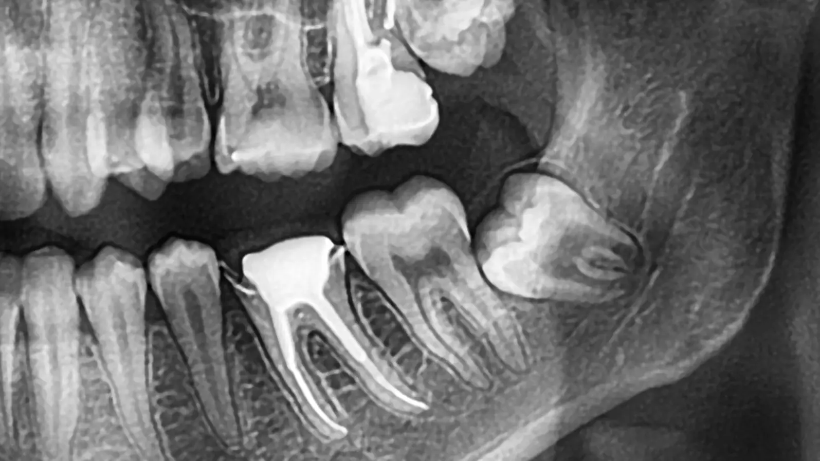
The analysis revealed that mandibular third molars that were quiet and healthy shared several distinct features on the X-ray, which could be identified with 68–87% certainty.
The four most common characteristics of a symptomless, pericoronitis-free mandibular third molar were:
Deep in the Bone: Most reliably predicted by location, 87% of symptomless teeth were deep in the bone, at or below the cementoenamel junction (the point where the crown meets the root) of the second molar, or only just perforating the cortex (classes A, B, and C).
Mesioangular Inclination: 75% of these teeth were tilted forward (mesioangularly inclined).
Reduced Bone Level: 70% were associated with a reduced marginal bone level behind the second molar. Surprisingly, the greater the bone reduction, the fewer the symptoms, which the researchers suggest may be related to the ongoing eruption process and natural bone remodeling in this young age group, rather than pathology.
Incomplete Root Development: 68% had roots that were not fully formed (incomplete root development). This is often tied to the tooth still being unerupted.
In addition, 78% of these quiet teeth were clinically unerupted (not visible in the mouth). Interestingly, signs of pathological changes in the follicle (the sac around the crown) were not a reliable predictor of a tooth's clinical status.
Practical Takeaway
These findings are clinically relevant for situations where a patient has an ambiguous infection and potential infection sources are being investigated, or when a dentist is interpreting an X-ray with limited clinical data.
By looking at a panoramic X-ray, clinicians can now use a profile of features—primarily a deep, mesioangularly tilted tooth with incomplete roots and reduced bone level—to exclude non-pathological mandibular third molars with a high degree of certainty, helping them focus on truly diseased teeth.
Article Details
Title: Radiographic identification of symptomless mandibular third molars without clinical pericoronitis
Authors: Tommi Vesala, Irja Ventä, Johanna Snäll, Marja Ekholm
Journal: Clinical Oral Investigations (2024) 28:561
DOI: 10.1007/s00784−024−05953−3