Oral Leukoplakia: Unraveling a Long-Standing Medical Mystery
Why one of the most common oral precancer conditions still puzzles scientists today
A Long-Known Disease That Remains Hard to Explain
Oral leukoplakia (OL) is one of the most common oral potentially malignant disorders—a group of oral conditions that may progress to cancer. Despite being recognized for nearly two centuries, experts still consider OL “enigmatic.”
Why does it appear in some people but not others?
Why do only certain lesions become cancerous?
And why is its diagnosis still inconsistent around the world?
These are the questions highlighted in a new narrative review by José Manuel Aguirre-Urizar, an expert in oral medicine and pathology, who revisits the historical, epidemiological, biological, and clinical complexities that make OL a persistent challenge.
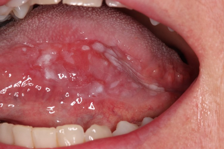
What Exactly Is Oral Leukoplakia? A Disorder Defined by What It Isn’t
Unlike most diseases, OL is defined more by exclusion: it is a white patch in the mouth that cannot be identified as any other condition.
This definition has evolved over decades, but the lack of a universally accepted description still leads to major variation in how OL is diagnosed and reported. In practice, this inconsistency makes it harder to track its prevalence, predict risks, or compare clinical outcomes.
How Common Is It? More Than We Think—But Estimates Vary Widely
Published studies report prevalence ranging from 0.33% to 11.74%, though an overall pooled estimate sits around 3.41%. Such wide variation is attributed to:
Differences in population demographics
Cultural habits such as tobacco, alcohol, or betel use
Genetic susceptibility
Variability in clinicians’ diagnostic criteria
Although previously thought to be a disease mainly affecting older male smokers, recent evidence shows that OL can also occur in women, younger adults, and even non-smokers.
What Causes It? A Multifactorial Condition With No Single Answer
The review emphasizes that the origins of OL are still unclear. Some cases remain idiopathic, just as first described in 1877. Known contributors include:
Tobacco and alcohol
Chronic irritation or trauma
Oral dysbiosis (imbalanced microbiome)
Chronic inflammation
Genetic and epigenetic changes
Researchers have identified several molecular alterations—such as deletions at 3p14 and 9p21, DNA methylation changes, and chromosomal instability—that may increase the likelihood of cancer progression.
Still, no biomarker has yet proven reliable enough for routine clinical use.
Diagnosing OL: More Complicated Than It Appears
Diagnosis requires both clinical examination and histopathological analysis:
Clinicians assess the appearance, location, and number of lesions.
Non-homogeneous types (e.g., nodular, verrucous, or erythroleukoplakic) carry a higher cancer risk.
Biopsy is essential—not only to confirm OL, but also to rule out look-alike conditions (e.g., candidiasis, lichen planus) and detect epithelial dysplasia.
Epithelial dysplasia—the degree of cellular abnormality—remains the strongest predictor of malignant transformation. However, grading dysplasia is still subjective, often leading to variability between pathologists.
Emerging technologies, such as AI-based histological assessment, show promise in improving consistency and predicting which lesions may progress to cancer.
How Often Does It Turn Into Cancer? Estimates Around 5–10%
Despite decades of research, studies report highly variable malignant transformation rates (from 0.09% to 38.5%).
Recent, more rigorous meta-analyses suggest a risk of 6–10%.
Key risk factors include:
Non-homogeneous clinical appearance
Lesions located on the tongue
Presence of epithelial dysplasia
Female sex and age >50 years (in some studies)
Larger lesion size
Tobacco use (significant in some, but not all analyses)
This variability again reflects inconsistent diagnostic criteria and follow-up strategies across studies.
Treatment: Still No Gold Standard
There is no universally agreed-upon treatment for oral leukoplakia. Key points from the review include:
Many medical therapies (vitamin A, retinoids, herbal extracts, etc.) show limited or inconsistent benefit.
Stopping tobacco may help some lesions resolve, but not reliably.
Surgical excision is commonly used—especially for high-grade dysplasia—but evidence that it prevents cancer is limited.
Even after complete removal, recurrence and malignant transformation can still occur.
One recent randomized clinical trial suggests surgery may reduce cancer risk in high-risk lesions, but further research is needed.
Given these uncertainties, lifelong follow-up is recommended for all patients, regardless of treatment choice.
Conclusion: A Call for Better, Standardized Research
After reviewing the available evidence, the author emphasizes one critical point:
We need well-designed, prospective clinical studies using clear, standardized diagnostic criteria.
Only through consistent diagnostic definitions, high-quality pathology assessments, and long-term follow-up can clinicians and scientists finally unravel the mysteries of:
why OL develops,
which lesions will become cancerous, and
how best to treat and prevent malignant transformation.
Oral leukoplakia has been known for nearly 200 years—but much about it still remains unsolved.
Original Article
Aguirre-Urizar JM. Oral leukoplakia: still an enigmatic disorder.
Med Oral Patol Oral Cir Bucal. 2025;30(5):e730–e735.
DOI: 10.4317/medoral.27214