Fighting Implant Infections: Why Killing Bacteria Isn’t Always Enough
How a new 3D laboratory model reveals the complex battle between biofilms, tissues, and antibacterial treatments
Dental implants have transformed modern dentistry, restoring chewing function and quality of life for millions of patients worldwide. Yet behind their high success rates lies a persistent challenge: peri-implant infections, driven by stubborn bacterial biofilms that are notoriously difficult to eliminate. A new study published in Scientific Reports takes a closer look at how common antibacterial agents interact not only with bacteria, but also with the surrounding oral tissues—using a highly advanced three-dimensional laboratory model.
The Hidden Problem of Biofilms Around Implants
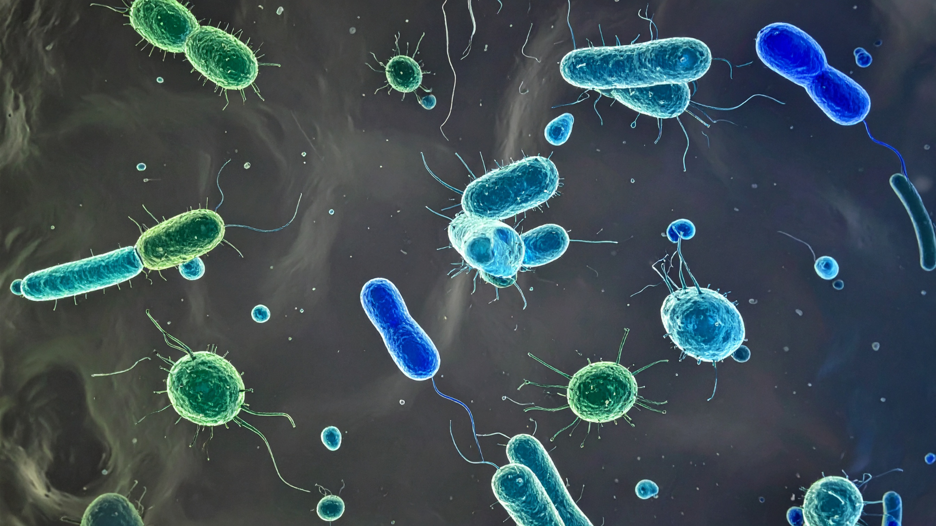
Unlike free-floating bacteria, microbes around dental implants often live in biofilms—dense, multi-species communities protected by a self-produced matrix. These biofilms can evade the immune system and resist antibiotics, making peri-implant mucositis and peri-implantitis difficult to manage clinically.
Traditional laboratory tests usually examine bacteria or human cells separately, often in flat (2D) cultures. While useful, these approaches fail to capture the real-life complexity of the mouth, where implant surfaces, oral tissues, and bacterial biofilms interact simultaneously.
A More Realistic Model: Recreating the Peri-Implant Environment
To overcome these limitations, the researchers used a sophisticated 3D system known as the INTERbACT model. This laboratory model combines:
A stratified human oral mucosa (epithelium and connective tissue),
A titanium dental implant,
A multispecies oral biofilm including Streptococcus oralis, Actinomyces naeslundii, Veillonella dispar, and the key pathogen Porphyromonas gingivalis.
Using this setup, the team tested several commonly used antibacterial agents: chlorhexidine, amoxicillin, ciprofloxacin, doxycycline, and metronidazole, applied at concentrations comparable to those found clinically around implants.
What Happens When Antibacterials Meet Biofilms?
The results revealed a nuanced picture.
While none of the antibacterial agents significantly reduced the overall size or structure of the biofilm, all of them increased the proportion of dead bacteria within it. In other words, the biofilms remained physically intact, but many of the bacteria inside were no longer viable.
Interestingly, the overall composition of the biofilm changed very little. Even P. gingivalis, a keystone pathogen strongly linked to peri-implant disease, largely survived across treatments. This finding highlights the remarkable resilience of mature biofilms and explains why infections can persist despite antibacterial therapy.
Protecting the Tissue Matters Too
Beyond bacterial killing, the study also examined how treatments affected peri-implant soft tissues. When biofilms were left untreated, clear damage to the epithelial layer was observed. In contrast, all antibacterial agents preserved tissue integrity, keeping the mucosa structurally intact and firmly attached to the implant surface.
This is an important insight: even if biofilms are not fully eradicated, antibacterial treatments can still help protect the soft tissue barrier, a critical defense against disease progression.
Not All Antibacterials Act the Same on Inflammation
Inflammation plays a central role in peri-implant disease, so the researchers measured inflammatory markers released by the tissues. Here, important differences emerged:
All treatments reduced hBD-2 and TIMP-1, markers associated with peri-implant inflammation.
Doxycycline significantly lowered the pro-inflammatory cytokines IL-1β and CCL20.
Chlorhexidine specifically reduced TNF-α, another key inflammatory mediator.