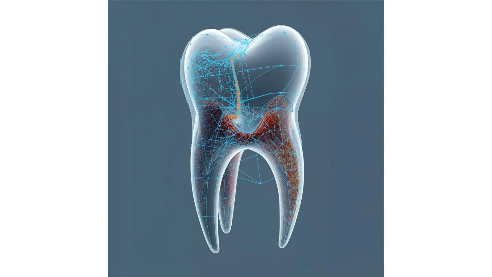
AI Maps Every Tooth in 3D: A Breakthrough for Dental Simulations
Dentists and orthodontists increasingly rely on 3D imaging to plan treatments — from braces to implants. A key step in this process is segmentation, or the digital separation of teeth, bone, and the tiny ligament space in between. Traditionally, this has been slow, labor-intensive work for specialists, who must manually trace every detail on cone-beam computed tomography (CBCT) scans.
Now, researchers at the University of Copenhagen and dental technology company 3Shape have developed an AI-based method that can do the job faster and with remarkable precision. Their approach not only identifies individual teeth and jawbone but also preserves the space for the periodontal ligament (PDL) — a soft tissue critical for accurate simulations of how teeth move under orthodontic forces.
Why the Periodontal Ligament Matters
In orthodontics, the PDL acts as a cushion and load-transfer zone between tooth and bone. If it’s missing from digital models, simulations used for planning treatments become unreliable. Many existing AI models fail to capture this delicate gap, resulting in anatomically inaccurate 3D reconstructions.
The new study set out to fix this problem: building a system that could automatically generate jaw models with realistic PDL spaces, while also distinguishing each tooth individually.
How the AI Works
The team adapted a known deep-learning architecture called MultiPlanar UNet (MPUNet). The process involved two main stages:
Pre-training – The AI first learned from “imperfect” datasets where tooth-bone gaps weren’t clearly marked.
Fine-tuning – It was then refined using a smaller set of highly accurate scans, guided by a special loss function that emphasized the importance of the tiny gaps where the PDL sits.
Finally, the researchers applied a marker-based watershed segmentation technique to separate neighboring teeth — a task often complicated by metal fillings, crowded teeth, or natural biting positions.
Promising Results
The AI was tested on 13 CBCT scans, including patients with implants, missing teeth, and image artifacts. Despite the challenges, the system consistently produced anatomically correct models suitable for finite element (FE) simulations — the same kind of biomechanical analysis engineers use to test stress and strain.
In fact, the method achieved a Dice score above 95% (a standard measure of segmentation accuracy) and showed superior performance in preserving gap regions compared to other state-of-the-art models. The resulting stress simulations on the PDL displayed smooth, realistic patterns, confirming that the models were ready for clinical and research use.
Why It Matters for Dentistry
By automating a task that usually requires hours of expert labor, this AI could speed up treatment planning, reduce costs, and increase accuracy. From orthodontics to implantology, more reliable digital models mean better predictions of how treatments will affect patients’ jaws.
The authors also highlight that their method works well even with limited high-quality training data — a common challenge in medical imaging. Future improvements could focus on training with more diverse scans or incorporating preprocessing to handle severe imaging artifacts.
Conclusion
This study shows that artificial intelligence can go beyond simple tooth detection: it can deliver highly detailed, biomechanically accurate 3D models of the human jaw. For dentists, orthodontists, and researchers, that means smarter simulations, fewer manual corrections, and ultimately, better patient care.
📖 Reference
Xu, P., Gholamalizadeh, T., Moshfeghifar, F., Darkner, S., & Erleben, K. (2023). Deep-Learning-Based Segmentation of Individual Tooth and Bone With Periodontal Ligament Interface Details for Simulation Purposes. IEEE Access.
DOI: 10.1109/ACCESS.2023.3317512