AI Learns to Map Dental Nerves With Unprecedented Precision
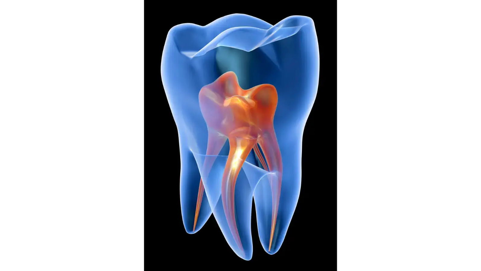
Why Mapping the Mandibular Canal Matters
When dentists plan dental implants, accuracy is everything. One of the greatest risks is damaging the mandibular canal — a delicate passage in the lower jaw that carries nerves and blood vessels. Traditionally, locating this canal relies on radiologists carefully marking 3D cone beam CT (CBCT) scans by hand. This process is not only time-consuming but also prone to human variability.
A New AI-Powered Approach
A team of researchers from Aalto University, Tampere University Hospital, and Planmeca Oy set out to change this with artificial intelligence. They developed a deep learning system based on a fully convolutional neural network to automatically identify the mandibular canal in CBCT scans.
Their dataset was impressive:
637 CBCT scans for training (with coarse expert annotations).
15 high-quality scans with voxel-level annotations for testing the model’s true accuracy.
This combination allowed the AI to “learn” from large-scale but imperfect data, then be tested against highly precise ground truth samples.
How Well Did It Perform?
The results were striking. The AI achieved an average error margin of just 0.5 mm, significantly outperforming earlier statistical shape-based models. In practical terms, this level of accuracy is more than sufficient for dental implant planning.
Interestingly, even though the training data contained rough, imperfect annotations, the deep learning model proved resilient to this “label noise” — it actually surpassed the quality of the coarse expert segmentations.
Beyond Accuracy: Speed and Efficiency
Automating mandibular canal segmentation could save radiologists hours of manual work, while also standardizing results across clinics. This has major implications for:
Implant dentistry – safer and faster surgical planning.
Maxillofacial surgery – better risk assessment before operations.
Future AI applications – such as bone density analysis and anatomical landmark detection.
Limitations and Next Steps
The model showed slightly reduced performance near the canal’s openings (the mandibular and mental foramina), where even human experts struggle due to anatomical complexity. Future studies aim to train the system on more diverse datasets, including patients of different ethnicities, imaging devices, and clinical variations.
Conclusion
This study demonstrates that AI is ready to assist — and even surpass — experts in one of dentistry’s most delicate imaging tasks. By reducing manual labor and increasing consistency, deep learning could soon become a standard tool in dental implantology and beyond.
Original Research Article
Jaskari, J., Sahlsten, J., Järnstedt, J., Mehtonen, H., Karhu, K., Sundqvist, O., Hietanen, A., Varjonen, V., Mattila, V., & Kaski, K. (2020). Deep Learning Method for Mandibular Canal Segmentation in Dental Cone Beam Computed Tomography Volumes. Scientific Reports, 10, 5842.