Hidden Architecture of Tooth Decay: Scientists Map How Bacteria Team Up to Destroy Enamel
How a tiny microbe, Streptococcus mutans, builds “corona-like” bacterial fortresses that trigger cavities
A New Look at an Old Foe
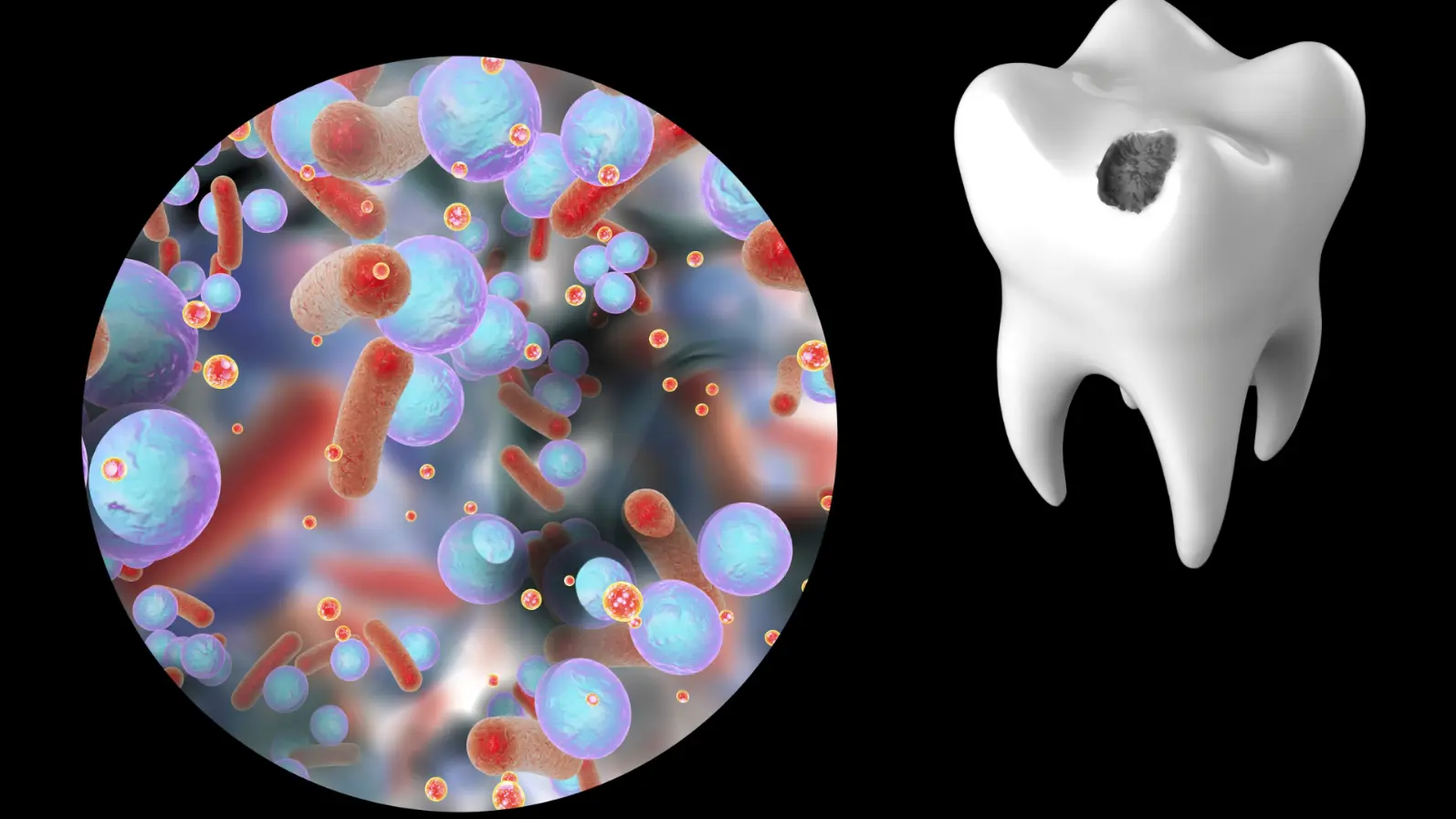
Tooth decay, or dental caries, affects more than 2 billion people worldwide. For decades, scientists have blamed one main culprit: Streptococcus mutans, a bacterium that thrives on sugar and produces acid that erodes tooth enamel.
But researchers from the University of Pennsylvania and Georgia Tech discovered that this bacterium doesn’t work alone it forms a precisely organized community of multiple species that act together to cause disease.
Using advanced 3D imaging and computational modeling, the team visualized intact biofilms from toddlers suffering severe cavities. Their findings, published in Proceedings of the National Academy of Sciences (PNAS), reveal a previously unseen “rotund” or dome-shaped bacterial architecture that resembles a tiny corona (crown)-like structure sitting on the tooth.
The Microbial “City” Behind Cavities
Inside these micro-communities, S. mutans occupies the inner core, while other oral bacteria such as Streptococcus oralis and non-mutans streptococci form outer protective layers.
This 3D organization isn’t random it’s actively built by S. mutans, which secretes an extracellular sticky scaffold (EPS glucan) that helps assemble the surrounding bacteria into this structured “city.”
This precise spatial organization, or biogeography, creates miniature acid-producing zones directly against the enamel surface. The team observed that these acidic microenvironments matched areas of localized enamel demineralization, confirming the community’s role in initiating caries.
Why Structure Matters
The architecture doesn’t just promote acid formation it also shields bacteria from antimicrobial agents and increases their resistance to harsh, low-pH environments.
When researchers disrupted the bacterial structure with enzymes that degrade the EPS matrix, the inner S. mutans cells became vulnerable to chlorhexidine (a common dental antiseptic) and showed reduced acid tolerance.
In essence, the “corona-shaped” biofilm acts like a protective bunker, allowing the core bacteria to survive treatments and maintain tooth-damaging activity.
Building the Fortress
To uncover how this architecture forms, the researchers grew mixed-species biofilms on human enamel in the lab. Over time, flat mixed communities transformed into dome-shaped clusters confirming that this structure develops progressively, guided by S. mutans’ genetic machinery. When the scientists used mutant S. mutans strains lacking the gtfB gene (responsible for producing the glucan scaffold), the bacteria failed to form the organized dome, and enamel damage was significantly reduced.
A New Way to Think About Cavities
This study changes how we understand tooth decay not as a single bacterium’s attack, but as the product of a coordinated microbial ecosystem.
The discovery of these highly ordered biofilm structures suggests that targeting the architecture not just the bacteri may offer a more effective way to prevent and treat cavities.
“Mapping the spatial structure of dental biofilms helps us see the ‘virulence hotspots’ where disease begins,” says lead author Dr. Hyun Koo. “By disrupting this organization, we might stop tooth decay before it starts.”
Reference
Kim, D., et al. (2020). Spatial mapping of polymicrobial communities reveals a precise biogeography associated with human dental caries. Proceedings of the National Academy of Sciences (PNAS), 117(22), 12375–12386.
DOI: 10.1073/pnas.1919099117